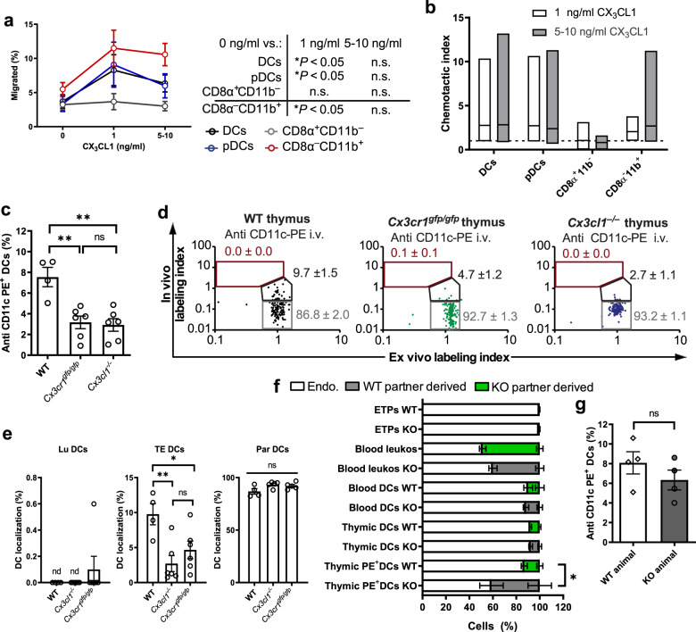
Fig. 6. Role of the CX3CR1-CX3CL1 pathway in thymic TE-DC positioning.
Thymic DC migration to gradients of CX3CL1 was assessed in transwell chemotaxis assays. Data are presented as a the percentage of migrated cells in each DC subset and b as a chemotactic index (ratio of the number of cells that migrated to media alone vs. media containing CX3CL1). n = 3 independent experiments with DCs from three to nine mice per group, two-way ANOVA. c–e Age and sex-matched C57B6 (WT), Cx3cr1gfp/gfp, and Cx3cl1–/– mice were injected with anti-CD11c-PE mAb IV and the thymus was harvested 2 min later to analyze c the frequency of PE+ DCs by FACS. n = 3 experiments, each with five mice/group (one-way ANOVA) and d intrathymic DC localization in each individual animal (representative examples are shown for each genotype) and e at a population level. n = 2 independent experiments, five mice/group (two-way ANOVA). f Leukocyte chimerism in blood and thymi of parabiotic Cx3cr1gfp/gfp (CD45.1+) and C57B6 (WT; CD45.2+) parabiotic mice. n = 7 pairs of mice (two-way ANOVA). g Parabiotic mice were injected with anti-CD11c-PE, euthanized after 2 min. and the frequency of PE+ cells among total thymic DCs was assessed in each partner. n = 5 pairs of mice (unpaired, two-tailed Student’s t test). Bars and error bars represent mean ± SEM. c **P = 0.0024 (WT vs. Cx3cr1gfp/gfp) and P = 0.0015 (WT vs. Cx3cl1−/−), n.s.: not significant. e **P = 0.0071, *P = 0.0475. f *P = 0.0356. DCs dendritic cells, pDCs plasmacytoid dendritic cells, Lu luminal dendritic cells, TE transendothelial dendritic cells, Par parenchymal dendritic cells. See also Supplementary Figs. 6 and 7.

