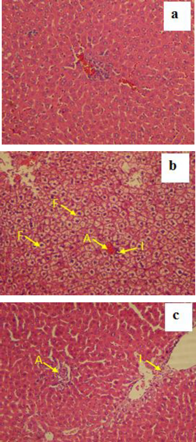
Figure 3.

Representative microscopic images (magnification X250) of H&E-stained liver sections following dust and clean air (CA) exposure accompanied by high-fat diet (HFD). a: HFD+clean air group showed normal appearance and there were no histopathological changes; b: The liver in HFD+N/S+ Dust exposed rats showed inflammation (I), accumulation of RBCs (A) and fatty deposit (F); and c: Animals in HFD+gallic acid + Dust group showed mild inflammation and blood cell accumulation but no signs of fatty deposit
