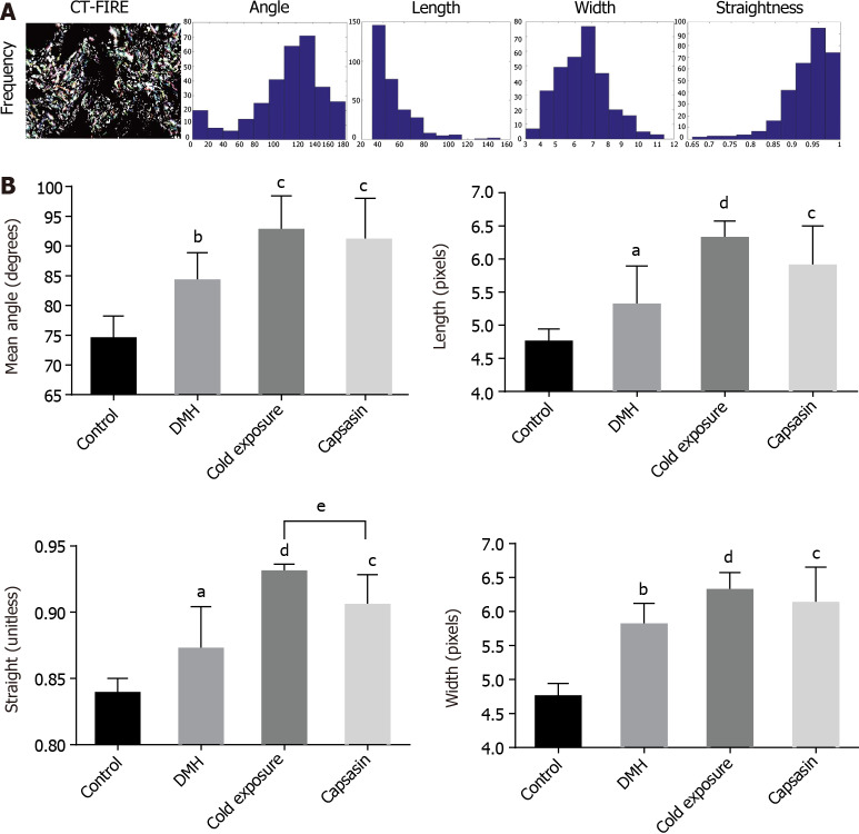
Figure 4.
Collagen fibers were automatically extracted for analysis using open-source software CT-FIRE. A: Histograms were generated to show the distribution of various parameters in each polarized light microscopy imaging; B: Quantitative analysis of collagen fibers from polarized light microscopy imaging in the colonic mucosa of different treatment groups. Data are mean ± SE of three images per tissues region. aP < 0.05, bP < 0.01, control compared with 1,2-dimethylhyrazine (DMH); cP < 0.05, dP < 0.01, DMH compared with cold exposure and capsaicin-treated group; eP < 0.05, Cold exposure compared with capsaicin-treated group.

