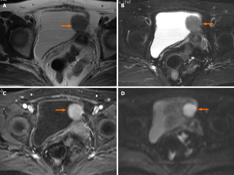
Figure 3.
Pre-operative contrast-enhanced pelvic magnetic resonance imaging. The orange arrow indicates a space-occupying lesion seen on the left wall of the bladder, originating from the bladder wall mucous membrane. It tended to infiltrate peripheral tissue, which was preliminarily suspected to be a desmoplastic fibroma or leiomyoma. A-D: It shows an oval structure that is isointense on T1WI (A) and T2WI (B) sequences; T1WI + fat suppression + enhanced sequence reveals noticeable enhancement (C); DWI sequence revealed limited diffusion (D).

