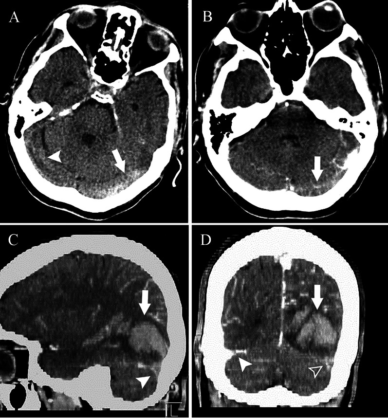
Fig. 1.
Brain computed tomography (BCT) of the patient. a Axial BCT without contrast material demonstrates stagnation in the left transverse sinus which appears hyperattenuated (cord sign) (arrow), while the right transverse sinus lumen is patent (arrowhead). b Axial BCT with contrast reveals filling defect in the left transverse sinus (arrow). c Sagittal BCT with contrast shows occipital lobe hemorrhagic venous infarct (arrow) along with empty delta sign and filling defect in the left transverse sinus (arrowhead). d Coronal BCT with contrast reveals the hematoma (arrow) as well as empty delta sign in the left transverse sinus (blank arrowhead) compared to normal contrast filling in the right transverse sinus (arrowhead)

