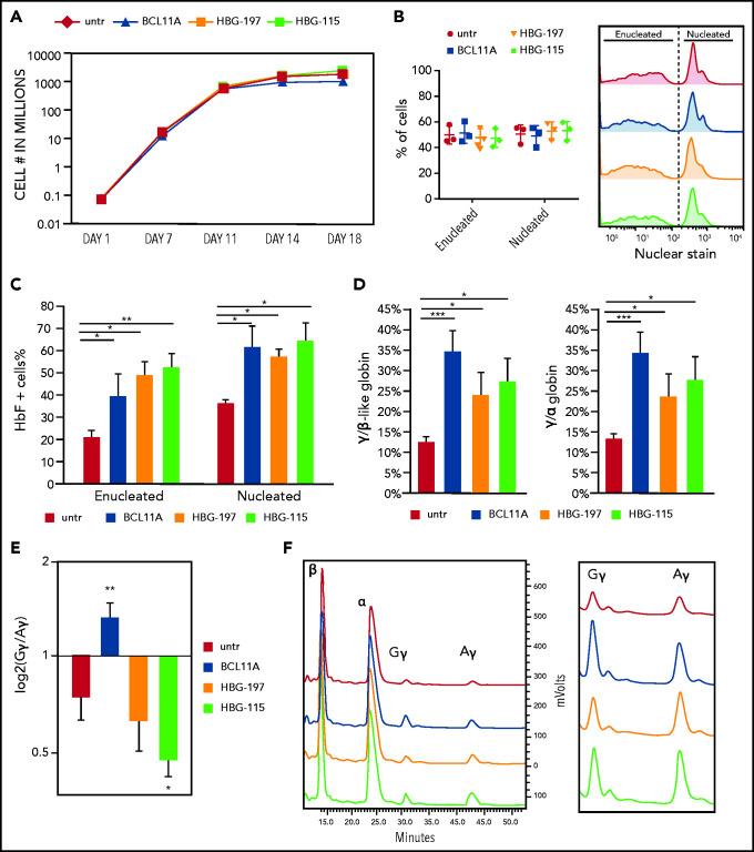
Figure 2.
Direct comparison of the 3 RNPs on their effect on erythropoiesis and HbF reactivation. (A) Cell growth over time during in vitro erythroid differentiation. (B) Enucleation of the erythroid cells on day 18 of the differentiation. (C) HbF+ cell frequency as assayed by flow cytometry. (D) HPLC analysis of γ-globin chain expression presented as ratio over the β-like globins and α-globin chain. (E) Ratio of Gγ/Aγ chains postediting with the 3 nucleases. (F) HPLC tracks depicting the difference in Gγ/Aγ balance between the different samples. All plots represent data from at least 4 different CD34+ cell donors. Values are represented as means ± SEM. ***P ≤ .0001, **P ≤ .001, *P ≤ .05 vs untreated (untr) (unpaired Student t test).

