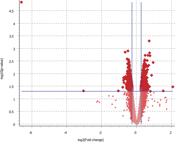
Fig. 1.
Volcano plot visualizing the statistical difference of the DEGs. The horizontal axis represents the p-value of detected transcripts, and the horizontal blue line at –log10 (P-value) of 1.3 is the threshold of 0.05. The vertical axis represents the fold-change (FC) of detected transcripts and the vertical blue lines are the cut-off criterion of 1.2. Each data point represents a gene or a variant of a gene. The upregulated and downregulated genes in the NEFAs treated group are illustrated as the dark red spots upper right and upper left, respectively

