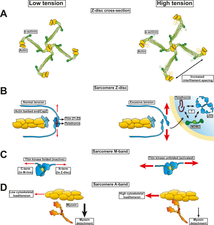
Fig. 2.
Striated muscle proteins showing structural deformation at rest compared to loaded in the sarcomere. (A) The α-actinin and actin lattice is deformed when muscles go from a relaxed to contracting state leading to an increased actin-actin separation. (B) Titin N-terminal domain anchoring at the Z-disc exhibits a unilateral resistance to increased loads in the direction of contractile force by thin and thick filament sliding. (C) The titin kinase domain of striated muscle unfolds with high tension from actomyosin contraction leading to autophosphorylation and activation of hypertrophic signaling pathways. With excessive loads telethonin leads to p53 downregulation possibly via direct interactions with p53 and its cognate E3 ligase MDM2. (D) Myosin detachment rate from actin post-power stroke decreases with increased loads against the direction of the myosin level arm swinging

