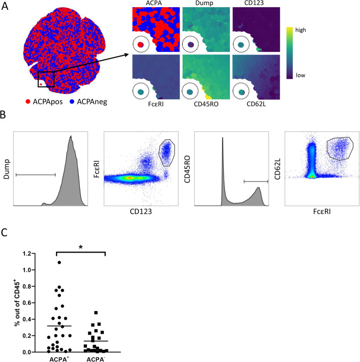
Fig. 3.
Independent flow cytometry replication confirmed the reduced presence of CD62L+ basophils in ACPA− samples. A Cytosplore dimension reduction of manually transformed flow FCS files of panel 1 showing both ACPA+ (red) and ACPA− (blue). Smaller panels on the right represent the cells within the backbox. Expression patterns matched that of MC cluster 18 (dashed circle), including CD123, FcεRI and CD45RO. CD62L expression was linked to ACPA status. B Gates used in Boolean gating in Flowjo to manually gate cluster 18. C Frequency of manually gated cells representing MC cluster 18. Subset frequency was lower in ACPA− RA (mean 0.32% vs 0.13%; p = 0.01)

