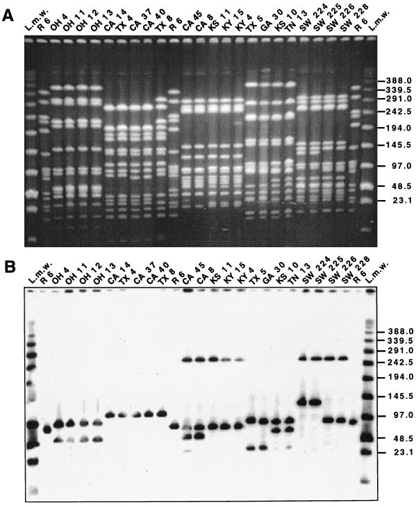
FIG. 1.
Association of PFGE pattern and lytA hybridization profile. Lanes marked L.m.w were loaded with low-range PFGE markers (New England Biolabs). Strain properties are listed in Table 1. SmaI fragments generated from the laboratory strain R6 served as additional molecular size standards. Numbers at right indicate molecular sizes in kilobases. (A) SmaI digest of total DNA separated by PFGE. (B) Hybridization of a Southern blot of the gel in panel A with the lytA gene probe.

