
FIG 11.
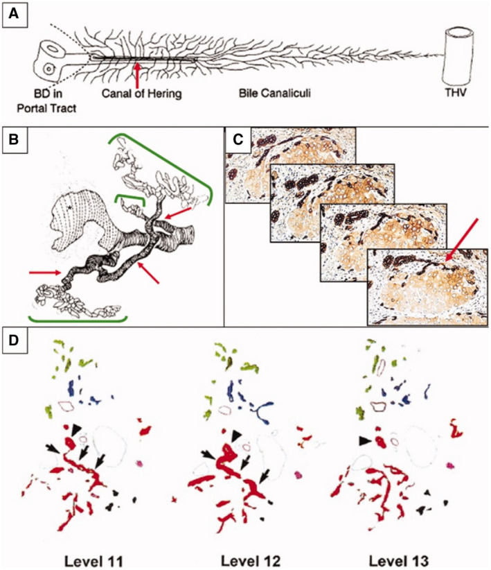
Relationship of DR hepatobiliary cells to the ductule/CoH stem cell niche. DR hepatobiliary cells are largely the “transit‐amplifying progeny” of intrabiliary stem cells. Their primary source is therefore the stem cell niche located in the most proximal branch of the biliary tree, the ductule/CoH unit. This relationship is best seen in 3D representations of DRs. (A) Schematic diagram of normal ductule and CoH (red arrow) structures and their relationship to BD, portal tract stroma, limiting plate, hepatocyte canalicular system (narrow, branching lines), and THV. Reproduced with permission from Hepatology. 3 Copyright 1999, American Association for the Study of Liver Diseases. (B) In primary biliary cholangitis, computer‐generated 3D reconstruction shows a granulomatous duct destructive lesion (large white area with vertical dashed black lines) and DR (green brackets) arising from preexisting ductule/CoH structures. Reproduced with permission from Journal of Pathology. 24 Copyright 1987, Pathological Society of Great Britain and Ireland. (C) Serial 4‐μm sections of hepatitis C virus–related cirrhosis. A small intraseptal hepatocyte nodule links to an interlobular BD via a single intermediate, K19‐positive, CoH‐like structure. The complete link can be appreciated only with examination of the serial sections. Immunostained with DAB; hematoxylin counterstain; original magnification, ×200. Reproduced with permission from Journal of Hepatology. 22 Copyright 2003, Elsevier. (D) Three sample tracings of K19‐positive DRs in sequential levels around a single portal tract. Colors are assigned to indicate contiguity of structures when analyzed in three dimensions. On each level, one BD is marked by an arrowhead. Note in levels 11 and 12 where the red arborizing structure connects via a single ductule/CoH branch to this BD (indicated by arrows). Reproduced with permission from Hepatology. 3 Copyright 1999, American Association for the Study of Liver Diseases.