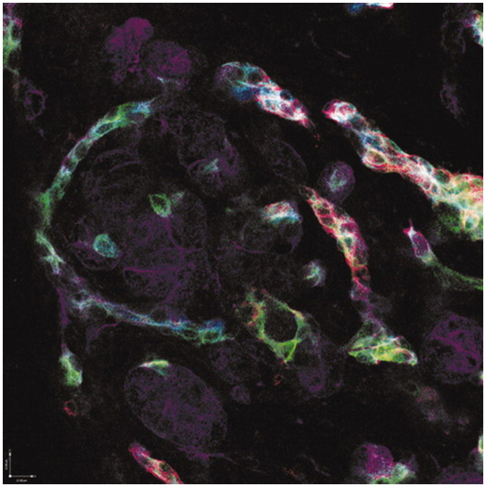
FIG 12.

Immunophenotypic diversity within DR hepatobiliary cells. Four‐color immunofluorescence of hepatitis C virus–associated DRs with K19 appearing as pseudocolored blue, K7 appearing as green, CD56 (neural cell adhesion molecule) appearing as red, and K18 (primarily staining hepatocytes) appearing as purple. Original magnification, ×200. Courtesy of E. Prakoso, N. Shackel, and G. McCaughan, University of Sydney, Sydney, Australia.
