
FIG 20.
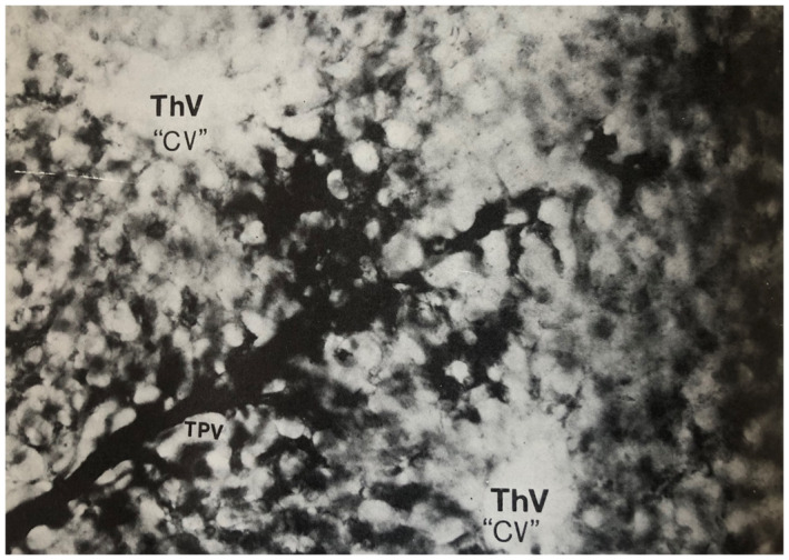
Human liver, simple acinus. The TPV branch of the structural unit is injected with Indian ink and runs perpendicular to the two ThVs or CVs with which it interdigitates. Comparison with figure 1.5 (reproduced as Fig. 17 in this manuscript) will indicate how the acinus occupies sectors of two adjacent classic lobules and extends between the two hepatic venule branches. Thick cleared section original magnification, ×270. Illustration originally by Prof. Aron Moses Rappaport, Toronto, and originally published in Pathology of the Liver. 40 Copyright 1979, Churchill Livingston. Reproduced with permission from the Publishing Editor.