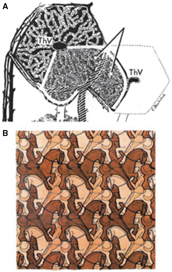
FIG 22.

(A) Horizontal section through a crosshatched Rappaport microvascular acinar unit of the liver (light gray, as indicated by the diverging lines), situated between two THVs (i.e., CVs), overlapping with the classic hepatic lobule shown in black. Reproduced with permission from The Anatomical Record. 41 Copyright 1954, American Association for Anatomy. (B) Horsemen: Woodcut in three colours by M.C. Escher, July 1946. M.C. Escher's “Horsemen, 1946” © 2021 The M.C. Escher Company‐The Netherlands. All rights reserved. www.mcescher.com.
