Figure 1.

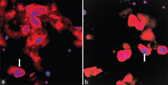
(a and b) Fluorescence imaging of cells prepared from voided urine. Cell morphology with arrow shows cell nucleus stained in blue and cytoplasm with orange fluorescence of TP4303 bound to VPAC receptors expressed on the cell membrane

(a and b) Fluorescence imaging of cells prepared from voided urine. Cell morphology with arrow shows cell nucleus stained in blue and cytoplasm with orange fluorescence of TP4303 bound to VPAC receptors expressed on the cell membrane