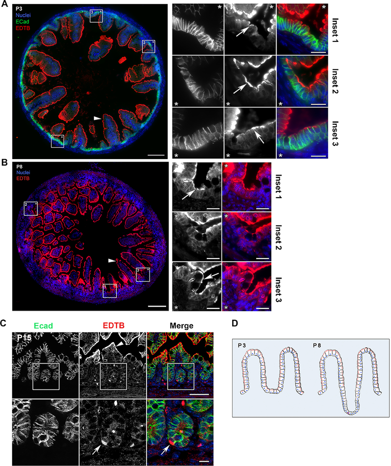
Figure 1. EDTB is localized to a subpopulation of cells in the intervillous and crypt regions.
(A-C) Ileum of P3, P8 and P15 mouse pups labeled with antibodies against E-cadherin (A, C, green) and EDTB (A, B, C, red). (A, B) Stitched images of the cross-sections show that EDTB is localized apically (arrowheads) along the length of the villi. Insets can be oriented by the placement of the asterisks. EDTB labels the intervillous region in P3 and P8 ileum (arrows; insets). Scale bar: 100μm, Scale bar inset: 20μm. (C) In P15 ileum, EDTB is localized apically within enterocytes along the villi (arrowhead) as well as in crypt cells (arrow), 6% of crypts contain an EDTB positive cell (n=117). Scale bar: 50μm, Scale bar inset: 10μm. (D). Summary of the distribution of EDTB expression during development. Before crypts form (P3), EDTB (red) is present in villous and intervillous enterocytes. When crypts begin to form, there are fewer EDTB-positive cells in the crypt, but there are occasional strongly labeled cells.

