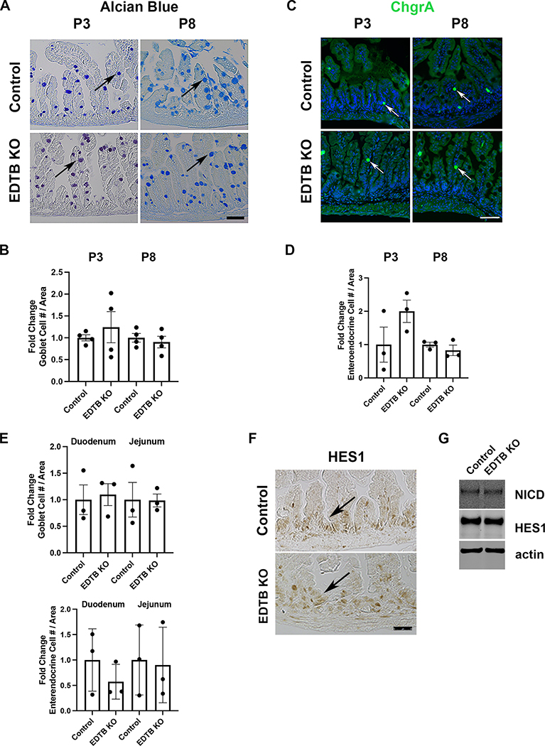
Figure 5. No change in intestinal secretory lineages with EDTB KO.
Ileum from P3, and P8 control and EDTB KO animals were stained for (A) Alcian blue (goblet cells)(arrows) or labeled with antibodies for (C) chromogranin A (enteroendocrine cells)(arrows) Scale bar: 50μm. (B,D). The total number of Goblet and Enteroendocrine cells per unit area was determined (n=3). There is no change in number of secretory cells in EDTB KO ileum compared to controls. (E) Duodenum and Jejunum from P8 control and EDTB KO were stained for Alcian blue or labeled with antibodies for ChgrA as above. There is no change in the number of secretory cells in the duodenum and jejunum in EDTB KO compared the control (n=3). (F) Ileum from P3 control and EDTB KO were labelled with antibodies for HES1. HES1 is localized to the nucleus in the intervillous region of control and EDTB KO ileum (arrows). (G) Lysates from P8 Ileum from EDTBfl/fl Cre− (control) and EDTBfl/fl Cre+ (EDTB KO) were examined by immunoblot for NICD and HES1 expression. NICD and HES1 expression levels are unchanged in EDTB KO ileum (n=4).

