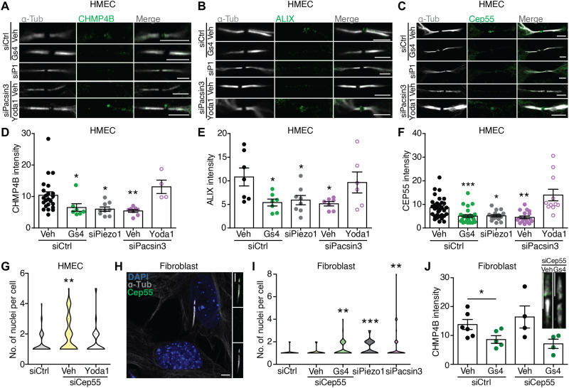
Fig. 3. Piezo1 regulates localization of ESCRT-III components to the late cytokinetic bridge.
Immunolocalization of α-tubulin and CHMP4B (A), ALIX (B), and Cep55 (C) at the midbody of siControl-, siPiezo1-, GsMTx4-, siPacsin3-, and siPacsin3 + Yoda1–treated HMECs. Scale bars, 5 μm. Quantification of CHMP4B (D), ALIX (E), and Cep55 (F) immunofluorescence intensities at the midbody. (G) Quantification of the number of nuclei per cell in HMECs transfected with control siRNA (n = 57) or siCep55 (vehicle = 73 and Yoda1 = 64) in the presence/absence of Yoda1. (H) Immunolocalization of Cep55 and α-tubulin in primary human dermal fibroblasts. Nuclear staining with DAPI. Scale bars, 5 μm. (I) Quantification of the number of nuclei per cell in primary human dermal fibroblasts transfected with control siRNA (n = 143), siCep55 (vehicle = 33 and GsMTx4 = 55), siPiezo1 (n = 61), or siPacsin3 (n = 57). (J) CHMP4B immunofluorescence intensities at the midbody of primary human dermal fibroblasts transfected with control siRNA or siCep55 in the presence or absence of GsMTx4. Scale bar, 5 μm. Data are means ± SEM. Number of cells (or experimental repeats) is indicated in each graph. Significance values are respect control condition as determined by Kruskal-Wallis test followed by Dunn’s post hoc test or ANOVA followed by Dunnett’s post hoc test (E).

