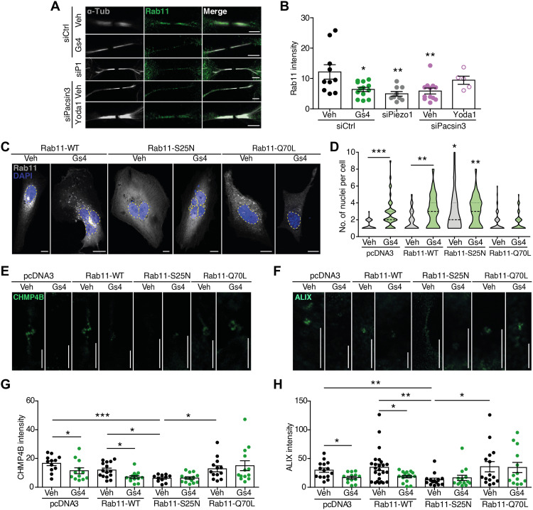
Fig. 4. Piezo1 controls delivery of Rab11-FIP3 endosomes to the cytokinetic ring.
(A) Immunolocalization of Rab11 at the midbody of siControl-, siPiezo1-, GsMTx4-, siPacsin3-, and siPacsin3 + Yoda1–treated HMECs. Immunolocalization of α-tubulin in white. Scale bars, 5 μm. (B) Quantification of Rab11 intensity at the ICB under the conditions is shown in (A). (C) Nuclear staining with DAPI (blue) in HMECs transfected with Rab11-WT-GFP, dominant-negative Rab11-S25N-GFP, or dominant-positive Rab11-Q70L–GFP in the presence/absence of GsMTx4. Scale bars, 10 μm. (D) Quantification of nuclei in HMECs overexpressing pcDNA3 (vehicle = 41 and GsMTx4 = 44), Rab11-WT (vehicle = 62 and GsMtx4 = 57), Rab11-S25N (vehicle = 50 and GsMTx4 = 38), or Rab11-Q70L (vehicle = 59 and GsMTx4 = 56) in the presence or absence of GsMTx4. CHMP4B (E) and ALIX (F) immunofluorescence intensities at the midbody of HMECs overexpressing pcDNA3, Rab11-WT, Rab11-S25N, and Rab11-Q70L in the presence or absence of GsMTx4. Scale bars, 5 μm. Quantification of CHMP4B (G) and ALIX (H) immunofluorescence intensities in HMECs under the conditions shown. Data are means ± SEM. Number of cells (or experimental repeats) is indicated in each graph. Significance values are respect control condition as determined by Kruskal-Wallis test followed by Dunn’s post hoc test or ANOVA followed by Dunnett’s post hoc test (B).

