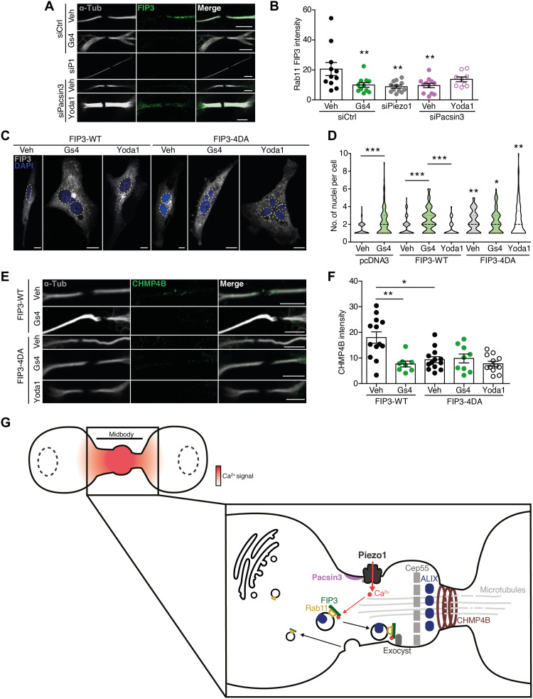
Fig. 5. FIP3 links Piezo1-generated Ca2+ signals to the recruitment of the abscission machinery.
(A) Immunolocalization of FIP3 at the midbody of siControl-, siPiezo1-, GsMTx4-, siPacsin3-, and siPacsin3 + Yoda1–treated HMECs. Immunolocalization of α-tubulin in white. Scale bars, 5 μm. (B) Quantification of FIP3 signal at the ICB under the conditions shown in (A). (C) DAPI staining of the nuclei in HMECs overexpressing FIP3-WT and FIP3-4DA in the presence and absence of GsMTx4 or Yoda1. Scale bars, 10 μm. (D) Quantification of nuclei in HMECs overexpressing pcDNA3 (vehicle = 97 and GsMTx4 = 104), FIP3-WT (vehicle = 114, GsMTx4 = 65, and Yoda1 = 71), and FIP3-4DA (vehicle = 75, GsMTx4 = 67, and Yoda1 = 89). (E) CHMP4B immunofluorescence of HMECs overexpressing FIP3-WT and FIP3-4DA in the presence and absence of GsMTx4 or Yoda1. (F) Quantification of CHMP4B immunofluorescence intensity in HMECs under the conditions shown. Data are means ± SEM. When required, number of cells (or experimental repeats) is indicated in each graph. Significance values are respect control condition as determined by Kruskal-Wallis test followed by Dunn’s post hoc test or ANOVA followed by Dunnett’s post hoc test (B). (G) Cartoon model of cytokinesis regulation by the Piezo1 channel. Mechanical forces exerted at the ICB activate Piezo1 channel generating a marked and localized increase in intracellular calcium (red shading). The increase in intracellular Ca2+ concentration is sensed by FIP3 to direct the transport of ALIX-containing Rab11-FIP3 endosomes to the ICB where ALIX recruits CHMP4B to complete abscission.

