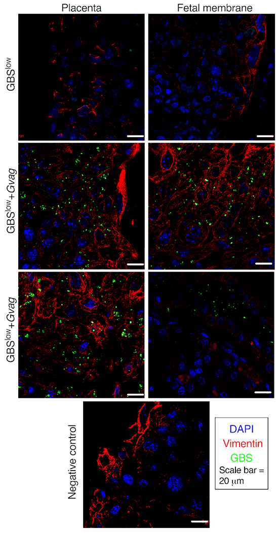
Figure 3. Co-inoculation with G. vaginalis facilitates GBS maternal-fetal unit invasion.

Representative images of immunofluorescence microscopy of the maternal-fetal interface from placentas isolated from dams inoculated with GBSlow or GBSlow+Gvag. GBS bacteria were detected with a monoclonal antibody (green). Sections were counterstained with DAPI (nuclei; blue) and vimentin (vasculature; red). Similar robust GBS staining was observed in placentas that were collected from GBSlow+Gvag dams that had placental infection evident by detectable cfu. The negative control panel (bottom) is a section from a GBSlow+Gvag placenta stained in parallel but omitting the GBS 1° antibody.
