. 2021 Oct 19;10:e69264. doi: 10.7554/eLife.69264

Figure 2. Morphological changes in pLL neuromast hair cells exposed to strong water wave stimulus.

(A) Schematic of a larval zebrafish. Blue dots indicate neuromasts of the lateral-line organs; green lines indicate innervating afferent lateral-line nerves. pLL neuromasts L3, L4, and L5 were analyzed (dashed circles). (B–C) Maximum intensity dorsal top-down 2D projections of confocal images of control or stimulus-exposed neuromast hair cells (blue (B) or orange (C); Parvalbumin immunolabel). Exposed neuromast hair-cell morphology was categorized as ‘normal’ i.e. radial hair-cell organization indistinguishable from control or ‘disrupted’ i.e. asymmetric organization with the hair-cell apical ends oriented posteriorly. (D) Maximum intensity projections of supporting cells (SCs) expressing GFP (green), immunolabeled synaptic ribbons (magenta; Ribeye b) and all cell nuclei (blue; DAPI). Note that SCs underlying displaced hair cells also appear physically disrupted (indicated by white arrows). Scale bars: 5 µm (F) Average percentage of neuromasts with ‘disrupted’ morphology following mechanical stimulation. Each dot represents the percentage of disrupted neuromasts (NM) in a single experimental trial. Disrupted hair-cell morphology was place dependent, with neuromasts more frequently disrupted following sustained stimulus and when localized toward the posterior end of the tail (*p = 0.0386, **p = 0.0049, ***p = 0.0004) (G) Average percentage of exposed neuromasts (NM) with ‘disrupted’ morphology in lhfpl5b mutants, which lack mechanotransduction specifically in lateral-line hair cells, vs. heterozygous WT. lhfpl5b mutants show a similar gradient of neuromast disruption following mechanical injury as WT siblings. Error Bars = SEM.

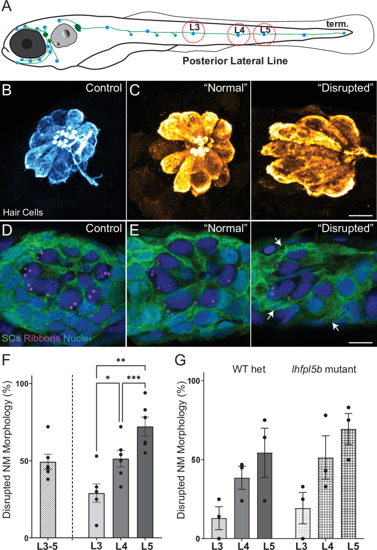
Figure 2—source data 1. Summary of normal and disrupted neuromast counts following sustained and periodic stimulus exposures.
Figure 2—source data 2. ummary of normal and disrupted neuromast counts in lhfpl5b mutants and wildtype siblings following sustained stimulus exposure.

Figure 2.

Figure 2—figure supplement 1. Fish exposed to periodic stimulus have less mechanical damage to neuromasts, but still show synapse loss.

Figure 2—figure supplement 1.

(A) Schematic of the two exposure protocols. Sustained exposure was a 20 min pulse followed by 120 min uninterrupted mechanical overstimulation; periodic exposure was 90 min exposure with intermittent 10 min breaks totaling 120 min. (B) Periodic stimulus causes less neuromast disruption. Immediately following sustained exposure, 46 % of exposed neuromasts showed a ’disrupted’ phenotype, whereas following a periodic exposure only 17 % of the neuromasts appeared ‘disrupted’ (Unpaired t-test **p = 0.0034). (C) Position of the neuromast along the tail was also associated with vulnerability to disruption with both sustained and periodic stimulation. (D-F’) Measurements of hair-cell number, innervation, and synapse number averaged from individual fish (neuromasts L3-L5). (D-D’) Average hair cell number per neuromast following exposure. With periodic exposure, the number of hair cells was comparable to control (*p = 0.108 sustained, p = 0.7233 periodic). (E-E’) % of neuromast hair cells innervated (****p < 0.0001 sustained, p = 0.0156 periodic). (F-F’) Average number of intact synapses per neuromast. There is significant loss of synapses with both periodic and sustained exposures (**p = 0.0045 sustained, *p = 0.0236 periodic).
Figure 2—figure supplement 2. Hair-cell organs of the ear appeared undamaged in larvae exposed to sustained stimulus.

Figure 2—figure supplement 2.

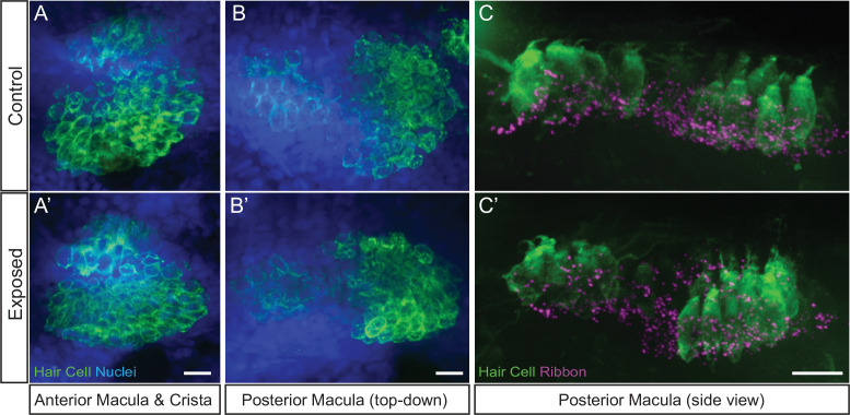
Representative maximum intensity images of hair cell organs in the ears of control (A–C) and larvae exposed to sustained strong water wave stimulus (A’)(-C’). Hair cells in A-B were immunolabeled with an antibody against Otoferlin; posterior macula in C were immunolabled with antibodies against Parvalbumin to label hair cells and CtBP to label synaptic ribbons. Scale bars: 10 µm.