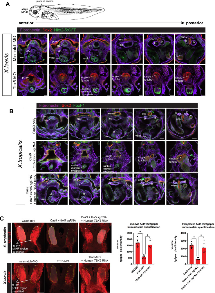
(A) Immunostaining of stage NF43 transgenic Xenopus laevis Tg(nkx2-5:GFP) reporter embryos injected with either mismatch-MO (control) or Tbx5-MO. Confocal optical sections through the cardiopulmonary-foregut region showing Fibronectin+ tissue boundaries (purple), GFP in the ventral pharynx, trachea, and ventricle; and Sox2 (red) in the pharyngeal/esophageal endoderm. Tbx5-depleted embryos exhibit severe cardiac hypoplasia, a lack of ventricular trabeculae, and a single, undivided foregut tube lacking Nkx2-1 respiratory progenitors. (B) Immunostaining of NF44 X. tropicalis embryos for Fibronectin (purple), Sox2 (red), and FoxF1 (green) shows that tbx5 mutants, have a single Sox2+ foregut tube and severe cardiac hypoplasia phenocopying X. laevis Tbx5 morphants. Injection of human TBX5 RNA rescues trachea-esophageal morphogenesis and cardiac chamber development. (C) Representative images for quantitation of Aldh1a2 immunofluorescence signal in Tbx5 LOF embryos at NF34. Nikon Elements Analysis AR software was used to determine the average volume pixel intensity of the fg lpm region (dotted yellow lines). Each dot in the graphs represents a distinct fg lpm/pSHF region from N=3 total embryos, all imaged with identical confocal laser settings. *p<0.05, parametric two-tailed paired t-test. at, atrium; eso, esophagus; lb, lung bud; LOF, loss-of- function; lpm, lateral plate mesoderm; MO, morpholino; pha, pharynx; tra, trachea; vent, ventricle.
Figure 2—figure supplement 1—source data 1. Quantitation of Xenopus NF34 fg lpm/pSHF Aldh1a2 immunostaining volume pixel intensity.


