. 2021 Oct 13;10:e69288. doi: 10.7554/eLife.69288

Figure 3. Tbx5 regulates Xenopus cardiopulmonary development in part via RA.

(A) Schematic of the experimental design. (B–J) Exogenous RA rescues Tbx5 LOF, while inhibition of RA phenocopies Tbx5 LOF. Whole-mount in-situ hybridization of NF34 X. laevis embryos after the indicated experimental treatments: injection of negative control 3 bp mismatch-MO (10 ng), Tbx5-MO (10 ng), Human TBX5 RNA (hTBX5; 100 pg), and/or 25 nM RA, 10 µM DEAB, DMSO vehicle control from NF20-34. The numbers of embryos with the observed expression pattern are indicated. Arrows indicate the relevant expression domain in the cardiopulmonar (CP) tissue. Brackets indicate the aSHF/pharyngeal domain. (K) Heat map showing relative expression from RT-PCR analysis of NF34 CP-foregut (fg) tissue dissected from control or Tbx5-MO injected embryos and treated with or without RA from NF20 to NF34. Each row is the average from the three biological replicates (n=4 explants per replicate). (L) Diagram of the proposed GRN model at NF25–35 showing the key role of Aldh1a2-dependent RA signaling downstream of Tbx5. White arrows indicate relationships tested in the above experiments and black arrows are demonstrated from the previous publications. Also see Figure 3—figure supplement 1, and related source data files. GRN, gene regulatory network; LOF, loss-of-function; MO, morpholino; RA, retinoic acid.

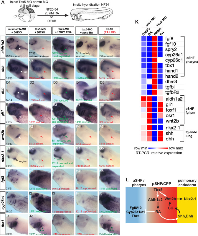
Figure 3—source data 1. Figure 3K.
Xenopus explant RT-qPCR source data. Explants were dissected at NF20, cultured 48 hr±DMSO or 25 nM all-trans retinoic acid (RA); harvested at NF34; three biological replicates for each condition; n=4 pooled explants in each replicate; pooled explants came from 2 to 3 separate fertilization/injection experiments.

Figure 3.

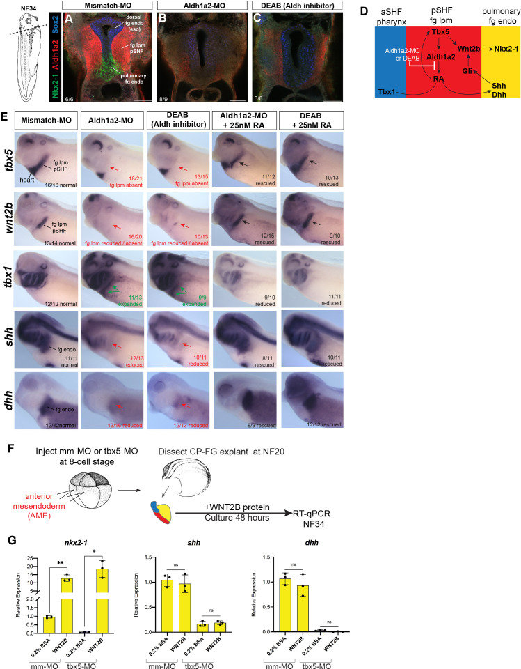
Figure 3—figure supplement 1. Aldh1a2 morpholino (MO) knockdown phenocopies DEAB treatment and WNT2B protein rescues Nkx2-1+ pulmonary fate.

Figure 3—figure supplement 1.

(A–E) Aldh1a2 MO knockdown phenocopies the pharmacological inhibition by DEAB treatment. Embryos were injected at the 8-cell stage into dorsal mesendoderm targeting the CP domain or treated from NF20 to NF34 with 10 µM DEAB or 25 nM RA. (A–C) Immunostaining of NF34 embryos for Aldh1a2 (red), Nkx2-1 (green), and Sox2 (blue) confirms that Aldh1a2-MO injection or DEAB treatment disrupts the RA>Tbx5>Aldh1a2>RA positive feedback loop (diagrammed in (D)) and results in failed induction of Nkx2-1+ pulmonary progenitors. (E) Whole-mount in-situ hybridization of NF34 control, Aldh1a2-MO injection, or DEAB treatment (NF20–34) showing similar effects. Treatment with 25 nM RA (NF20–34) rescued the changes in gene expression. The numbers of embryos with the observed expression pattern are indicated. Arrows indicate the relevant expression domain in the foregut – cardiopulmonary region. (F) Experimental schematic. (G) WNT2B protein 100 ng/ml rescues nkx2-1 but not Hh ligand expression in Tbx5-depleted fg explants. Graphs show mean relative expression ± standard deviation from N=3 biological replicates (four explants/replicate). Each black dot in the graphs represents a biological replicate (pool of n=4 explants). *p<0.05, **p<0.01, parametric two-tailed paired t-test relative to uninjected, untreated explants. CP, cardiopulmonary; ns, not significant; RA, retinoic acid.
Figure 3—figure supplement 1—source data 1. Xenopus explant RT-qPCR source data.
Exogenous WNT2B protein treatment rescues nkx2-1 expression in Tbx5-depleted Xenopus cardiopulmonary foregut explants. RT-qPCR analysis of Xenopus foregut cardiopulmonary explants treated ± recombinant Human WNT2B protein (100 ng/ml) 48 hr NF20–34.