. 2021 Oct 29;10:e67858. doi: 10.7554/eLife.67858

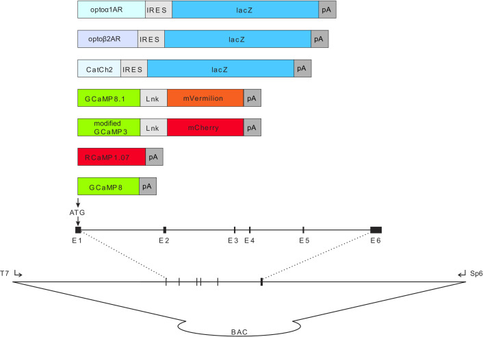
Figure 1. Combinatorial optogenetic mice.

The figure illustrates the general paradigm of tissue-specific cellular activation and simultaneous response decoding with tissue-specific optical sensors. Here, a biallelic SA nodal CatCh2/cardiomyocyte-GCaMP8 mouse is generated from a single cross of isogenic (C57BL/6J), hemi-allelic effector/sensor mice, as in Figure 2H. Mice with specific expression of optoeffectors (cardiac conducting tissue, cardiac and smooth muscle syncytia, endothelium, sympathetic neurons, T cells, and glia) and optosensors (SA nodal tissue, cardiac and smooth muscle, endothelium, ciliated epithelium, and alveolar epithelium) are reported. LA: left atrium; RA: right atrium; RV: right ventricle; LV: left ventricle; Ao: aorta; SVC: superior vena cava; SAN: sinoatrial node.

© 2021, Virginia Greene

Figure adapted from commissioned work by Virginia Greene. This figure is not available under the Creative Commons Attribution License and commercial use requires permission from the copyright holder.

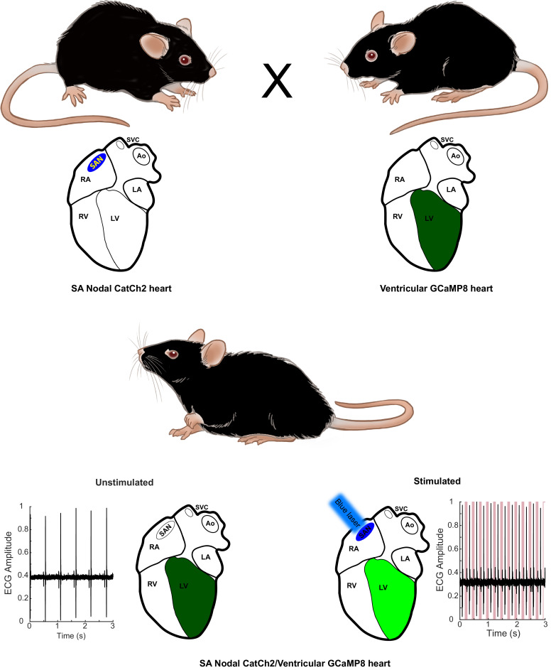
Figure 1.

Figure 1—figure supplement 1. Diagram of expression cassettes used in construction of CHROMus mice listed in Table 1.

Figure 1—figure supplement 1.

Transgene expression cassettes are placed in frame with the ATG initiation codon from the desired target gene in the desired BAC molecule using standard BAC recombineering protocols. The optogenetic transgene cassettes contain the coding sequence for the optogenetic protein (optoα1AR, optoß2AR, or CatCh2) and the bacterial LacZ coding sequence separated by an internal ribosome entry sequence (IRES) element. Expression cassettes for fluorescent reporter proteins contain circularly permutated GFP or RFP coding sequence flanked by synthetic M13 peptide and calmodulin coding sequences, 5′ and 3′, respectively. In some cassettes, a fusion protein was created by linking an RFP (mCherry or mVermilion) to the carboxy terminus of the GCaMP protein separated by a spacer peptide. The purified recombinant BACs were microinjected into single-cell zygotes at Cornell University or UC-Irvine as described in Materials and methods.