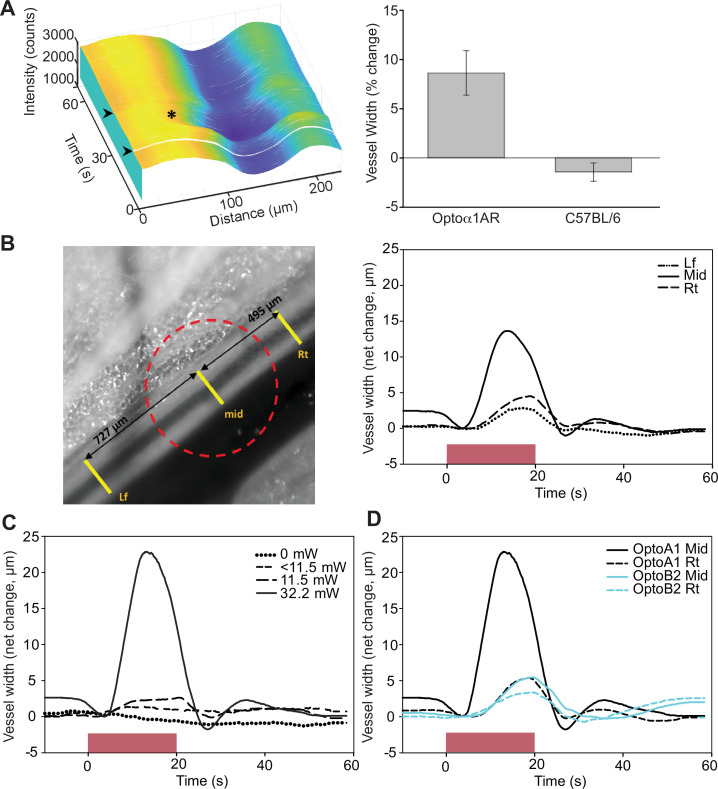
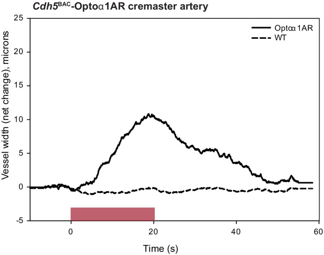
Figure 7. Endothelial cell optoeffector function.
(A) Laser stimulation of femoral artery in Cdh5BAC-Optoα1AR induces vasodilation. Left panel: MATLAB-generated 3D rendering of vessel width and intensity over time. The arrowheads mark the start and end of laser activation, and the asterisk marks dilation of the artery. Right panel: comparison of peak change in vessel width between Cdh5BAC-Optoα1AR_IRES_lacZ and C57BL/6J control mice. ***p=0.003, Holm–Sidak (± SE). Optoα1AR data were derived from five femoral arteries from five animals, and the C57BL/6J data were derived from five femoral arteries from three animals. Source data: Figure 7—source data 1. (B) Activation of Optoα1AR protein results in migration of vasodilation away from the laser irradiation point. Left panel: brightfield image of the femoral artery with positions of the mid, right (Rt), and left (Lf) analysis positions indicated. Red dashed circle indicates laser irradiation spot on the vessel. Right panel: change in vessel width over time. Source data: Figure 7—source data 2. (C) Vessel relaxation is dependent on laser intensity. Source data: Figure 7—source data 3. (D) Comparison of vasodilation induced by Optoα1AR and Optoß2AR proteins with laser power at 32.2 mW. Source data: Figure 7—source data 4. The data in (B–D) represent experiments with three animals.


