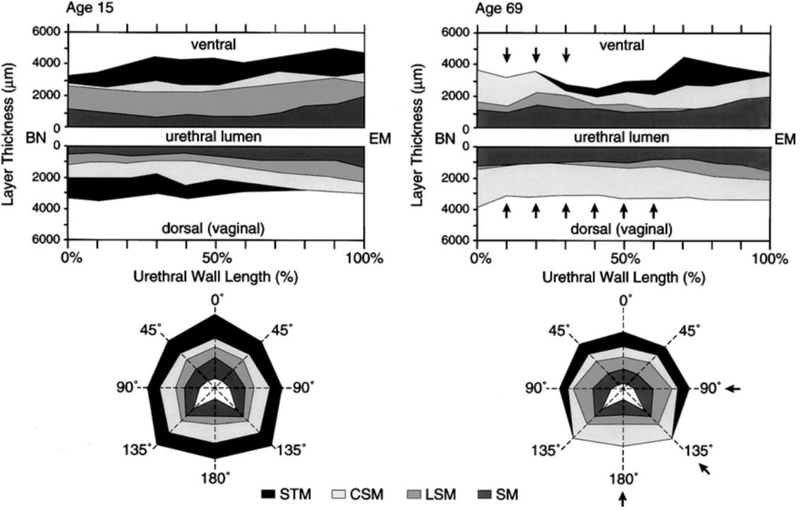
Figure 5.
Graphic illustration of the muscle loss pattern of urethral wall with aging based on histologic findings. Upper plots show the layer thickness in mid-sagittal section. Lower plots are the corresponding mid-urethral cross-sections. Note the proximal loss (arrows) in the thickness of the striated muscle in both the dorsal wall and the mid-urethral cross-section with age. BN, Bladder neck. EM, external meatus. STM, Striated muscle. CSM, Circular smooth muscle. LSM, Longitudinal smooth muscle. SM, Submucosa (from Perucchini, 2002).

