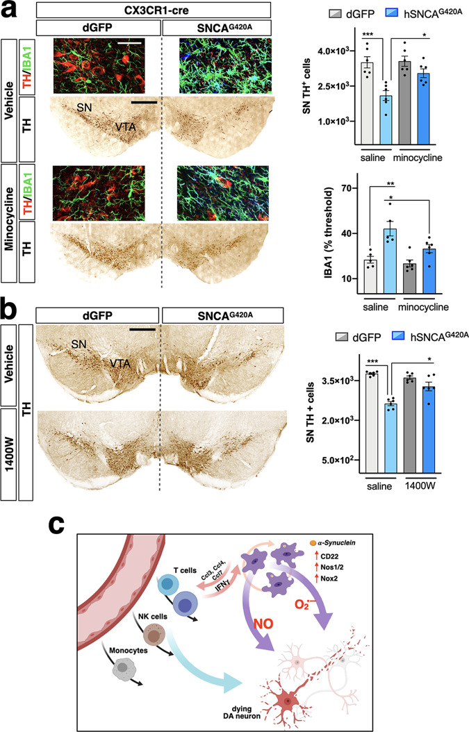
Fig. 8. Chronic administration of minocycline and 1400 W attenuates neurodegeneration in CX3CR1-SNCA mice.
a Chronic minocycline administration significantly enhances survival of nigral DA neurons in CX3CR1-SNCA mice 5 weeks after viral transduction. αSYN microglial accumulation accounts for 40% ± 1.8% of DA neuronal cell loss compared to the control side, which is significantly rescued by the minocycline treatment (a, upper bar graph). The cell loss is accompanied by the ~2-fold increase of IBA1 signal compared to control side, which is attenuated by the minocycline treatment (a, lower bar graph). Data are the mean ± s.e.m from n = 6 nigral tissues. Two-way ANOVA is followed by Bonferroni’s post-test. Upper bar graph: ***p = 0.0009, *p = 0.03; lower bar graph: **p = 0.001, *p = 0.04. Scale bar, 45 µm (fluorescence micrographs), 300 µm (IHC micrographs). b CX3CR1-SNCA mice treated with the iNOS inhibitor 1400 W showed a strong recovery in DA neuronal survival 5 weeks after viral transduction. Microglial expression of αSYN accounts for 28% ± 0.45% of dopaminergic cell loss compared to the control side, which is rescued by the 1400 W administration. Data are the mean ± s.e.m from n = 6 nigral samples. Two-way ANOVA is followed by Bonferroni’s post-test. ***p < 0.001, *p = 0.014. Scale bar, 300 µm. c Illustration of the mechanisms underlying DA neuronal degeneration by αSYN accumulation in microglia. αSYN-dependent exhausted microglia upregulate chemotactic molecules to recruit peripheral immune cells. Infiltrated immune cells and αSYN accumulation synergize in establishing a vicious circle that fuel massive release of oxidant molecules and subsequent DA neuronal cell loss. SN substantia nigra, VTA ventral tegmental area.

