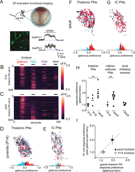
Figure 2. Disproportionate representation of glabrous skin emerges in the brainstem over postnatal development.
A. (top) A preparation allowing in vivo multiphoton imaging of the gracile nucleus (GN) of the brainstem. (bottom) A representative, motion-corrected image of thalamic PNs expressing jGCaMP7, and corresponding raw fluorescence time series extracted from ROIs centered on two example neurons (black, 100 μm scale bar) and surrounding neuropil fluorescence (gray) during skin stroke.
B. Touch-evoked responses of thalamic projection neurons functionally imaged in the GN. Calcium signals were evoked by stroking equivalent skin areas of the hindpaw glabrous skin (red), hindpaw hairy skin (blue), thigh (purple), and back (black) in P14 mice, data displayed is pooled across n=3 mice (100 μm scale bar).
C. Representative response to touch of the body in adult mice, similar to B.
D. Representative map of the body within thalamic projection neurons of the GN. A glabrous skin preference index, (glabrous − hairy) / (glabrous + hairy), was computed for all neurons responding to hindpaw skin stimulation in this and other experiments (bottom). At P14, there are roughly equal proportions of glabrous preferring and hairy preferring cells.
E. Representative map of the body within inferior collicular projection neurons in the GN, similar to D.
F-G. Representative maps of the body in adulthood for thalamic projecting neurons (F) and inferior collicular projecting neurons (G).
H. The proportion of thalamic or inferior collicular projecting neurons in the GN that preferentially respond to hindpaw glabrous skin touch expands over developmental time (p < 0.01, Mann-Whitney U test).
I. The relationship between glabrous preference in the GN of the brainstem and glabrous preference in hindpaw S1 at two developmental timepoints.

