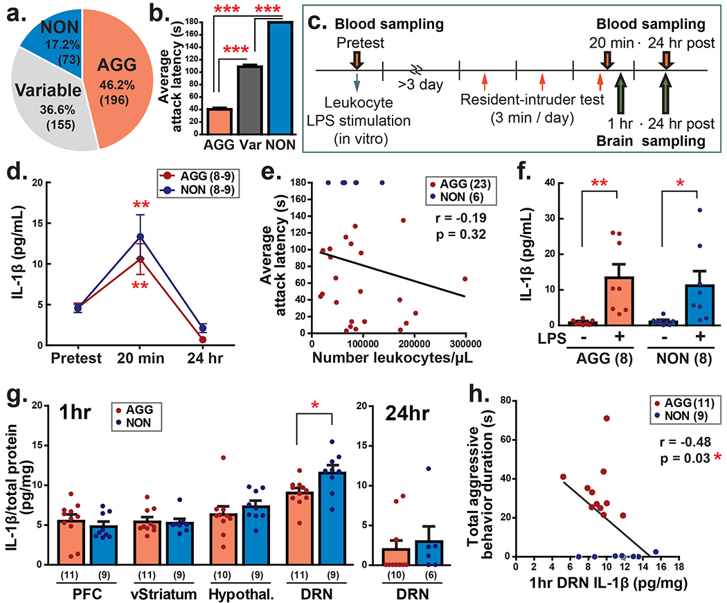
Figure 1. Individual difference of aggressive behavior and IL-1β response in the periphery and central nervous system.

a Ratio of aggressors (AGG), non-aggressors (NON), and variable aggression animals (Variable) in 424 resident CD-1 male mice. b Average attack latency over 3 days of resident-intruder encounters in AGG, Variable (Var) and NON animals (right). c Schematic timeline of experiments. d Blood IL-1β level before, 20 min, and 24 hrs after the aggressive encounter in AGG and NON animals. Dunnet’s t-test was conducted in each group to compare 20 min and 24 hrs samples with the base level (pre). e IL-1β production from cultured leukocytes by LPS stimulation in AGG and NON. f There was no correlation between number of leukocytes and average attack latency over 3 days of resident-intruder test. g IL-1β level in the prefrontal cortex (PFC), ventral striatum (vStriatum), hypothalamus (hypothal.), and dorsal raphe nucleus (DRN) 1 hour after the aggressive encounter (Left). IL-1β level in the DRN decreased 24 hours after aggressive encounter (right). h Negative correlation was observed between duration of total aggressive behaviors and IL-1β level in the DRN 1 hour after the aggressive encounter. Numbers in the parenthesis indicate the number of animals in each group. *p<0.05, **p<0.01, ***p<0.001.
