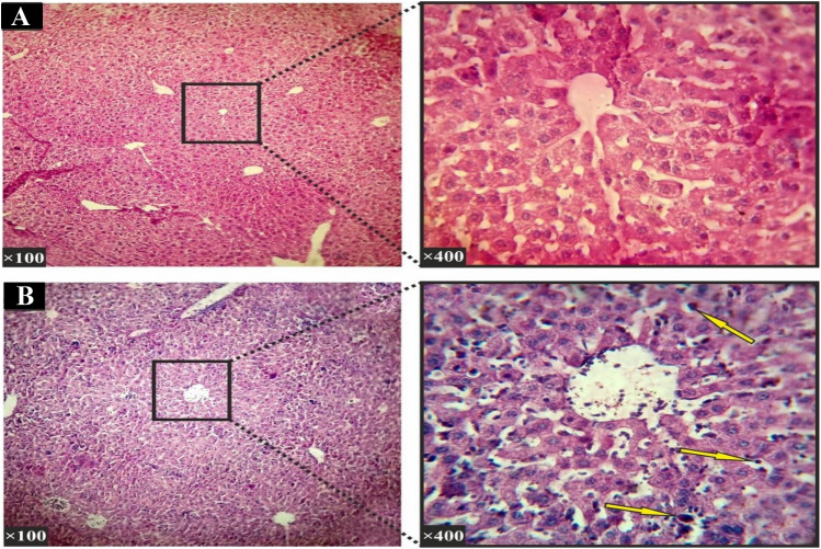
Fig. 6.
Histopathological section of the liver (Hematoxylin–Eosin staining). a The negative control group: In the histological study of the liver in the control group, Hepatocytes, their dispersion and liver sinusoids were normal and there was no hyperemia, edema and swelling. b A sample section for the group received 400 mg/kg: The study of liver histopathological sections showed visible inflammatory cells in the liver parenchyma (shown with arrows)

