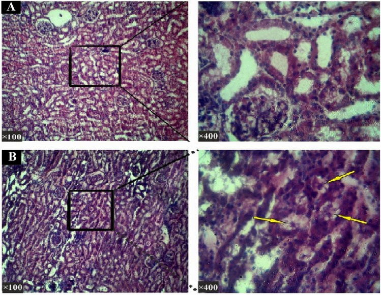
Fig. 7.
Histopathological section of the kidneys (Hematoxylin—Eosin staining). a In the histological study in the negative control group of the kidneys in the control group, renal corpuscle, proximal and distal tubules and the collecting ducts were normal. b In the group received 400 mg/kg dosage: The kidney histopathological studies of the sections in this group showed visible degradation in the lumen of the proximal and distal tubules. The cell wall of the proximal and distal tubules seen to be peeling off and dropping into the inner lumen (shown with arrows) and renal parenchyma disrupted too

