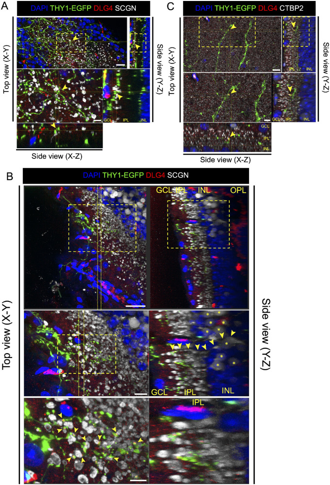
Figure 5.
Presumptive synapse formation between transplanted THY1-EGFP+ cells and host bipolar cells after transplantation. (A) Four weeks after transplantation in the control eye, three-dimensional confocal images of transplanted area stained with postsynaptic marker DLG4 (red) and cone bipolar cell marker SCGN (white). DLG4 was co-localized at the contact site (yellow arrows) of THY1-EGFP+ cells (green) and host bipolar cells. Scale bars: 20 µm (upper panel) and 6 µm (lower panel). (B) Four weeks after transplantation in the NMDA-injected eye, three-dimensional confocal images of transplanted area stained with postsynaptic marker DLG4 (red) and cone bipolar cell marker SCGN (white). DLG4 was co-localized at the contact site of THY1-EGFP+ cells (green) and host bipolar cells (host bipolar cell bodies marked by yellow asterisks). A bipolar cell axon (yellow arrows) can be traced back from the IPL into the INL of the host retina. Scale bars: 30 µm (upper panel), 10 µm (middle panel), and 6 µm (lower panel). (C) Four weeks after transplantation in NMDA-injected eye, three-dimensional confocal images of transplanted area stained with postsynaptic marker DLG4 (red) and presynaptic marker CTBP2 (cyan). DLG4 was co-localized at the contact site (yellow arrows) of THY1-EGFP+ cells (green) and CTBP2. Scale bars: 20 µm (upper panel) and 5 µm (lower panel).

