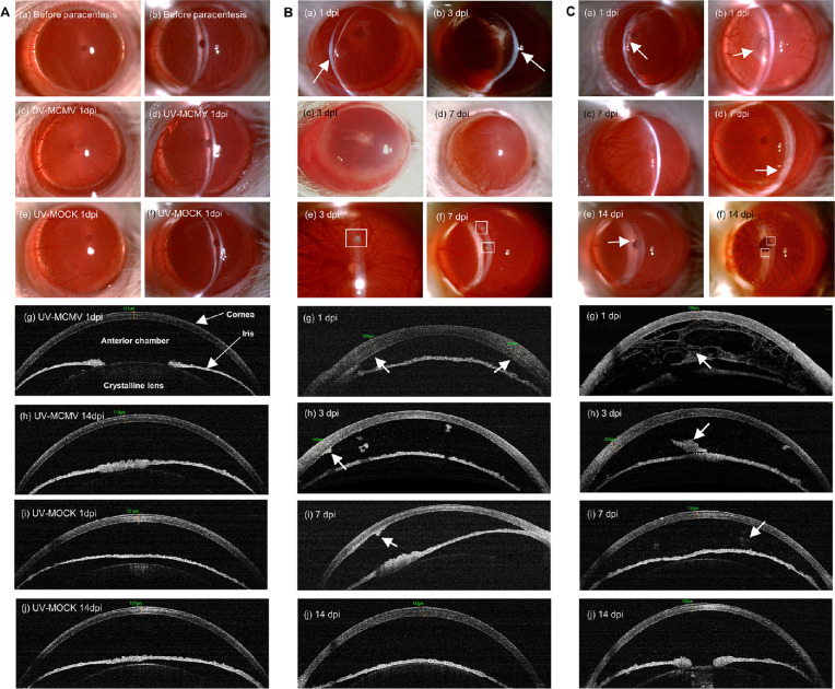
Figure 2.
Clinical manifestations of CMV keratouveitis in rats by slit-lamp examination and AS-OCT. (A) Slit-lamp views and AS-OCT images of the anterior segment of the eyes in different control groups. (B) Representative images of corneal lesions in the MCMV group: (a, b) Localized corneal edema (white arrows); (c, d) Diffuse corneal edema; (e, f) Mutton fat KPs (white squares); (g) OCT view of uneven corneal edema (white arrows); (h, i) KPs exhibiting “hump”-like structures with high reflectivity by OCT (white arrow); (j) Corneal edema and anterior chamber inflammation relieved at 14 dpi. (C) Representative images of anterior uveitis in the MCMV group: (a) Diffuse anterior chamber exudates (white arrow); (b) Occlusion of pupil (white arrow); (c) Posterior synechia with exudate in the pupil area; (d) Whitish appearance of the inferior iris (white arrow); (e, f) Iris nodules (white arrows); (g) OCT view of “net”-like anterior chamber exudates (white arrow); (h) Exudative membrane around the pupil on OCT (white arrow); (i, j) Residual anterior chamber exudates exhibited sporadic patches (white arrow) or fine dots with high reflectivity on OCT. Original magnification of slit-lamp images, × 25 magnification; AS-OCT, anterior segment optical coherence tomography; dpi, days postinfection.

