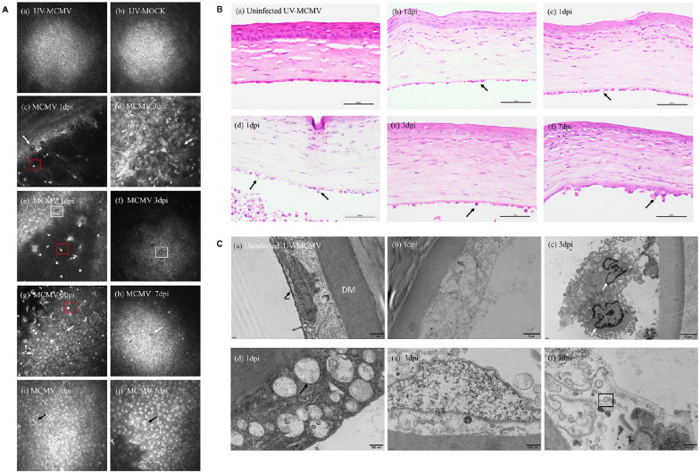
Figure 4.
Imaging and histopathological changes in the corneal endothelium after infection. (A) IVCM image of the corneal lesions: (a, b) Normal corneal endothelium; (c, d) “Black hole” changes (white arrows); (e) Enlarged intercellular gaps (white square); (f) Distinctive endothelial cell with “high reflection” nucleus surrounded by a dark ring area (white square); (g, h) Inflammatory cellular infiltration (white arrows); (I, j) Endothelial cells with bright nucleus or merged nuclei (black arrows). (B) Representative results of endothelial damage by H&E staining: (a) Normal corneal structure; (b) Swelling of corneal endothelial cells (black arrow); (c) Ballooning degeneration (black arrow); (d) Cell rupture with inflammatory cell infiltration (black arrows); (e, f) Neutrophils and monocytes attached to the injured endothelium (black arrows). Bar = 50 µm. (C) TEM view of the endothelial layers: (a) Normal endothelial cells full of mitochondria (black arrow); (b) Cell disruption; (c) Endothelium destroyed and infiltrated by monocytes (white arrows); (d) Severe swelling of mitochondria (black arrow); (e) Nuclear edema with mitochondrial damage; (f) Viral envelope within the cytoplasm (black square). IVCM, in vivo confocal microscopy; TEM, transmission electron microscopy; dpi, days postinfection.

