Figure 6.
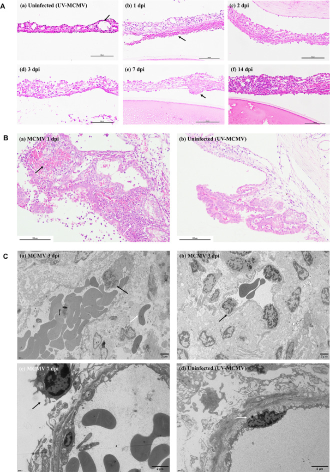
Pathological changes in the iris and ciliary body based on light microscopy and TEM. (A) Histological examination of the iris at different postinfection times by H&E staining: (a) Normal blood vessel of the iris (black arrow); (b) Leakage of red cells and exudation of inflammatory cells and fibrins due to the disruption of the blood-aqueous barrier (black arrow); (c, d) Iris edema and infiltration of inflammatory cells; (e) Fibrous membrane on the iris (black arrow); (f) Thickened iris with lymphocyte infiltration on 14 dpi. Bar = 100 µm. (B) Representative image of ciliary body lesions after MCMV infection: (a) Vascular congestion (black arrow) with large numbers of neutrophils infiltrating into the stroma (white arrow); (b) Normal ciliary body of rats in the UV-MCMV group. Bar = 100 µm. (C) TEM view of the iris after infection: (a, b) Migrating monocytes that deformed and adhered to the vascular wall (black arrows), red cell leakage through the disrupted vascular wall (white arrow); (c) Monocyte infiltration (black arrow); (d) Normal vascular endothelial cell of the iris.

