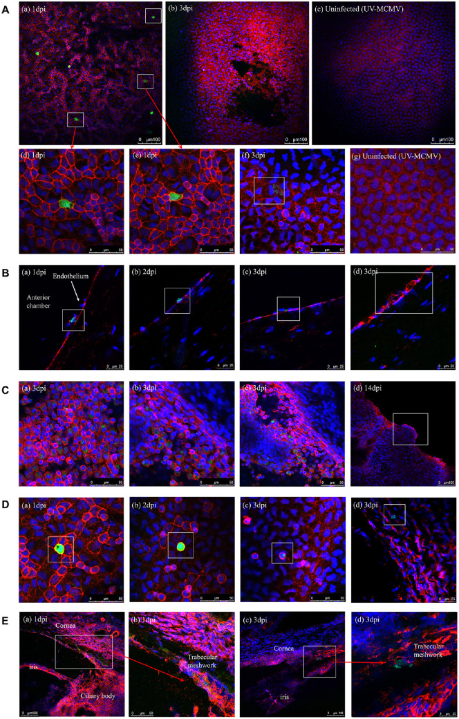
Figure 7.
MCMV infection site detection by immunofluorescence examination. (A) Infection sites of MCMV in the endothelial layer: (a) Multiple infected endothelial cells expressing green fluorescence at 1 dpi (white squares); (b) Patchy necrosis and detachment of endothelium noted at 3 dpi; (c) Endothelial layer without signs of infection in the UV-MCMV group; (d, e) Enlarged images of infected endothelial cells with white squares in picture-a; (f) Swelled endothelial cells with mild green signals within the cytoplasm (white square); (g) Uninfected normal endothelium. (B) Identification of MCMV infection sites on frozen sections: (a) Infected endothelial cells interacting with two inflammatory cells identified by blue nuclear markers (white square); (b, c) Infected endothelial cells; (d) Injured endothelial cells with fractured membranes. (C) Infection sites of MCMV in the iris: (a–c) Infected iris cells expressing green fluorescence within the cytoplasm; (d) Hyperplasia of the margin of the pupil representing the iris nodule. (D) Infected monocytes. (E) Infected TM cells in the frozen sections.

