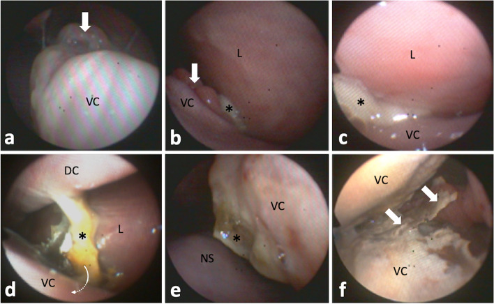
Fig. 2.
Left sinonasal endoscopic views. a Polypoid mucosal swelling (arrow) of caudodorsomedial ventral conchal (VC) aspects. Endoscope is located in the middle nasal meatus laying dorsal on the VC looking caudal. b Polypoid mucosal swelling (arrow) of dorsolateral VC aspects near the rostral nasomaxillary aperture, which shows draining purulent secretions (asterisk). The endoscope was guided towards the left lateral wall of the nasal cavity (L). c Advancing the endoscope further into the aperture clarified origin of purulent secretions (asterisk) from the rostral maxillary sinus, which was not accessible due to mucosal swelling. d At the level of entrance to the VC recess (arrow), masses of isolated necrotic conchal bone and impacted pus (asterisk) are visible. The VC recess was not accessible endoscopically. Dorsal concha, DC. e Caudomedial aspects of the deformed VC showing inflamed, partially necrotic, areas with purulent deposits (asterisk). Nasal septum, NS. f VC sinus reached via a large necrotic defect in middle medial aspect of the VC. Isolated necrotic conchal bone an pus (arrows) hindered passage into the rostral maxillary sinus

