Fig. 3.
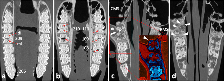
Coronal CT image sections of maxillary cheek teeth and adjacent sinonasal structures. a – d Section plane moving in apical direction, starting 10 mm subocclusal (tooth 209); 0.6 mm slice thickness; W3100/C500. a Subocclusal enlargement and filling with hypodense material of the mesial infundibulum (mI) of 209; the distal infundibulum (dI) appears unaltered. b At the infundibular fundus level, mI 209 appears highly enlarged and filled with hypodense material and gas; dI appears unaltered. The respective mI of 108, 109, 110, and both infundibula of 210 show accentuated signs of cement hypoplasia and hypodense/ gas filling. c Normal pneumatization of caudal maxillary sinuses (CMS) and right rostral maxillary sinus (RMS); soft tissue/ fluid dense filling of left RMS (asterisk). Insert: enhanced visualization of thin sinusoidal mucosal lining at the air interface and normal appearing delicate sinusoidal lamina dura of the apically unaltered neighboring tooth 210 (arrowhead); Perfusion Color Look Up Table (CLUT), soft tissue = red, bone = dark blue. Osteolytic loss of sinusoidal lamina dura of the buccodistal root of 209 resulting in an apicosinusoidal fistula (arrow). Partial loss of interradicular bone (plus) and widening of periapical periodontal space (open arrowheads). d Severe sclerosis of interalveolar and periapical alveolar bone of 209 (arrowheads). Inflammation caused discontinuity in the nasal lamina dura of the palatal root, thus apiconasal fistulation (arrow)

