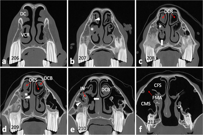
Fig. 6.
Transversal CT image sections of the head. a–f Section plane moving from rostral to caudal; 0.6 mm slice thickness; W3100/C500. a Dorsal concha (DC) and ventral concha with its ventral conchal recess (VCR) appear unaltered at the level of 106/206. b Ventral conchal deformation and disintegrated VCR entrance area (asterisk) at the level of interdental space 07/08. Soft tissue/ fluid dense filling of left dorsal conchal recess (arrowhead). c Ventral conchal deformation (asterisk) and retraction (arrow), and filling of the left dorsal conchal recess (DCR) at the level of IDS 08/09. Right DCR appears pneumatized (normal). d Soft tissue/ fluid dense filling of the left ventral conchal sinus (asterisk) and VCR (arrowhead), dorsal conchal bullae (DCB), and left dorsal conchal sinus (DCS) at the level of 109/209. e At the level of IDS 10/11 the medial aspect of the ventral concha appears with a large necrotic defect (arrow). The mucosa of the rostral maxillary sinus (arrowhead) and around the infraorbital nerve (IN) appears swollen. f At the level of 111/211 the caudal maxillary sinus (CMS) and conchofrontal sinus (CFS), which communicate via the frontomaxillary aperture (FMA), appear unaltered

