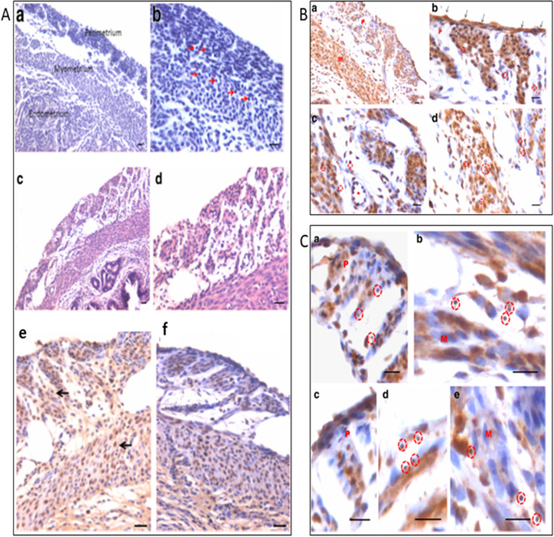
Fig. 17.
Uterine myometrium in adult mice after 5 days of FSH treatment to bilaterally ovariectomized mice. A a&b: Myometrium of bilaterally ovariectomized mice with small spherical stem cells (red arrows) c, d: After 5 days of FSH treatment e,f: PCNA expression in FSH treated group. B a-d: OCT-4 expression in FSH treated myometrial sections. Note presence of small, spherical cells expressing FSHR and OCT-4. C a-d: Higher magnification showing small spherical cells with nuclear OCT-4. Note bigger myometrial ‘progenitor’ cells with cytoplasmic OCT-4 [67]. Scale: 20 μm

