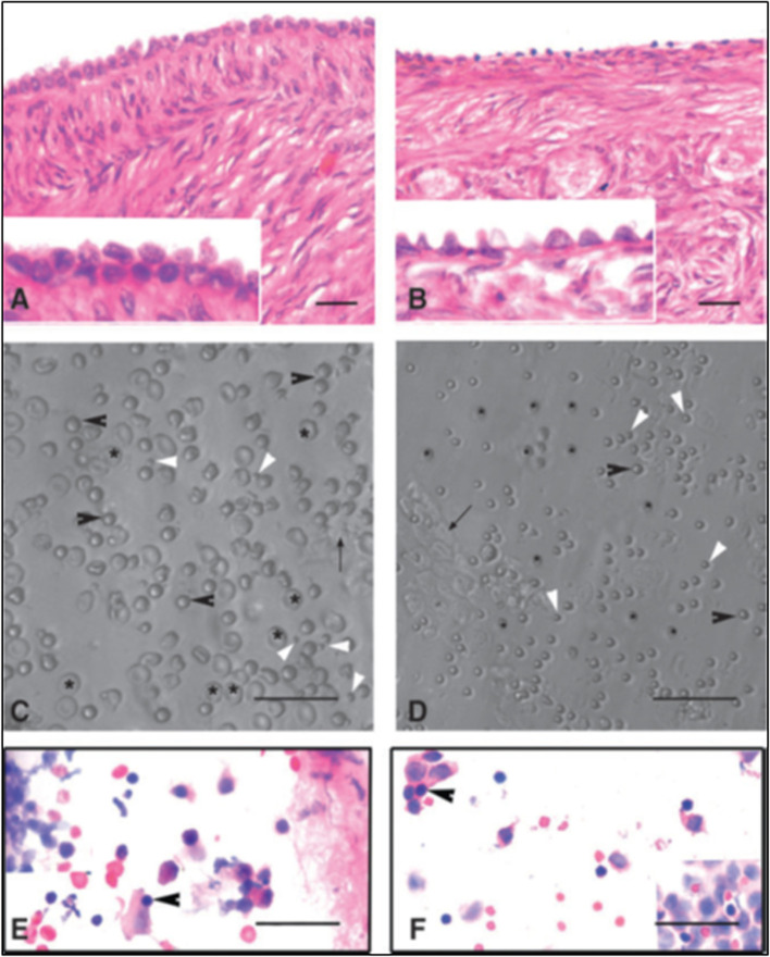
Fig. 2.
Stem cells reside in adult ovary surface epithelium (OSE) A& B. H&E stained sections of menopausal human (left panel) and sheep (right panel) ovarian cortex. Note prominent OSE cells which were scraped and collected in a dish. C& D. Note the presence of stem cells under Hoffman optics amongst OSE cells including small VSELs (white arrowhead), OSCs (black arrowhead) along with red blood cells (asterix). Epithelial cells with pale stained nuclei and abundant pink cytoplasm were also observed E & F. Darkly stained, spherical stem cells in H&E stained cell smears were clearly visualized (arrowheads) [23]. Scale: 20 μm

