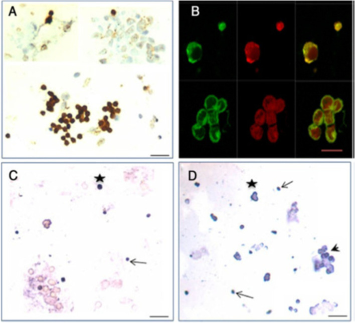
Fig. 3.
FSHR expression on ovarian stem cells. A. Immuno-localization studies on OSE cell smears shows that only the stem cells express FSHR and epithelial cells remain distinctly negative. B. Immunofluorescence studies localized FSHR in small VSELs, slightly bigger OSCs and in a cluster of cells. In situ hybridization results using specific oligo probes showed expression of (C) Fshr-1 and (D) Fshr-3 transcripts on FSH treated sheep OSE smears. As evident Fshr-1 transcripts were observed in the nuclei of both VSELs (arrow) and OGSCs (asterix). Fshr-3 transcripts were localized both in the nuclei and cytoplasm and even in the germ cell nests ‘cysts’. Presence of Fshr-3 in both cytoplasm and nuclei suggested active involvement of Fshr-3 transcript during FSH action on the stem cells [24]. Scale: 20 μm

