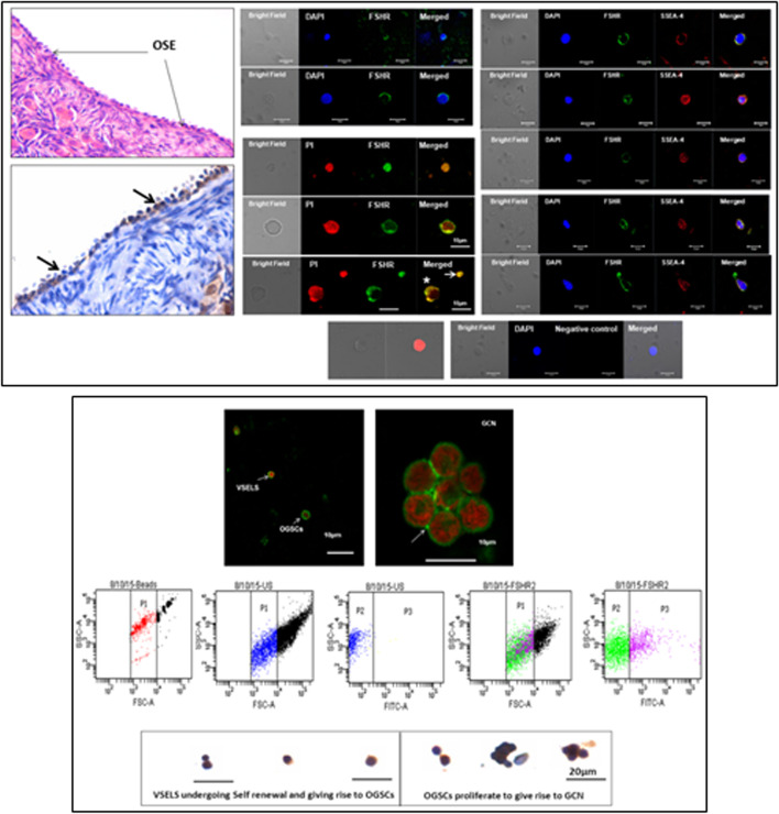
Fig. 4.
Upper panel: Sheep ovarian sections on the left clearly show FSHR expression in the OSE. Stem cells isolated from sheep OSE expressed FSHR. Co-expression of FSHR with SSEA-4 (cell surface marker for pluripotent stem cells) was clearly evident confirming FSHR expression on the stem cells. Lower panel: FSHR expression on VSELs and OSCs and on a germ cell nest (multiple cells cluster with cytoplasmic continuity). 2–6 μm cells were studied for FSHR expression by flow cytometry. Asymmetric, symmetric divisions and clonal expansion of stem cells is shown [24, 25]

