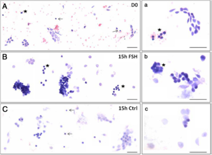
Fig. 5.
Effect of FSH treatment on the ovarian stem cells located in the ovary surface epithelium. A&a Freshly prepared sheep OSE smear after H & E staining. Epithelial cells (spindle-shaped cells with pale nuclei and abundant cytoplasm) and distinct populations of darkly-stained putative stem cells including the VSELs (arrow) and OSCs (asterisk) were evident along with red blood cells (RBCs) B&b Stem cells and germ cell ‘cysts’ were increased after 15 h of FSH treatment. The nests represent clonal expansion of stem cells with incomplete cytokinesis C&c OSE smear after 15 h culture without FSH. Scale: 20 μm [24]

