
Figure 2.
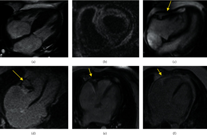
CMR findings in a patient with CA and CP. (a) Four-chamber cine function shows LV hypertrophy and thickened IAS in a CA patient. (b) Short-axis LGE depicts transmural LV and RV GD enhancement in CA. (c) Four-chamber cine function shows pericardial thickening (yellow arrow) in a CP patient. (d) LGE image in four-chamber view shows significant localized pericardial thickening and calcification (yellow arrow). (e, f) Localizer and LGE images of a 49-year-old woman with a history of shortness of breath and palpitations from three months ago. The patient had restrictive physiology on echocardiography. For further evaluation, CMR was performed, which showed restrictive physiology with normal pericardial thickness (arrow), and notably, a moderate reduction in the strain values (GLS: −14.01%), which was more in favor of CP. Hemodynamic finding in invasive angiography was an indicator of constriction. Finally, CP with normal pericardial thickness was confirmed during surgery.