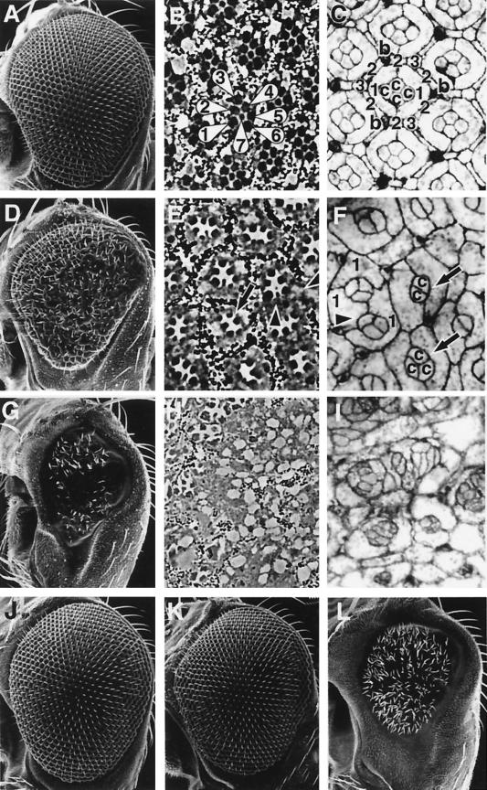
FIG. 1.
Phenotypes induced by ectopic TAK1 signaling. Scanning electron micrographs of the compound eye (A, D, G, J, K, and L), tangential histological sections (B, E, and H), and pupal eyes (at 40 h after puparium formation) stained with cobalt sulfide (C, F, and I) are shown. (A to C) Canton-S. (A) The wild-type eye is composed of a regular array of about 800 ommatidia. (B) Each ommatidium contains six outer photoreceptor cells and an inner photoreceptor cell, R7 (R1 to R7 are indicated with numbers). (C) Cobalt sulfide staining shows the apical profile of cells in the epithelium. Each ommatidium contains four cone cells (indicated with “c”) and two primary pigment cells (“1”), surrounded by six secondary pigment cells (“2”), three tertiary pigment cells (“3”), and three interommatidial bristles (“b”). (D to F) pGMR-mTAK overexpression. (E) Most of the ommatidia contain only five to six photoreceptors (arrow), and visual pigments are also disrupted at many positions (arrowheads). (F) In the pupal eye, most of the ommatidia contain only two or three cone cells (arrows) and occasionally are also missing primary pigment cells (arrowhead). Numerous numbers of interommatidial cells are also missing in this mutant. (G to I) pGMR-mTAK1ΔN overexpression. (G) Expression of mTAK1ΔN in the developing eye results in a severe decrease in size of the compound eyes. (H and I) Ommatidial structures are totally disrupted and hard to discriminate in the adult head section and pupal eye disc. (J) pGMR-hTAB1. Expression of hTAB1 alone in the eye has no effect on eye development. (K) Weak pGMR-mTAK1 overexpression phenotype. (L) Coexpression of hTAB1 and mTAK1 (weak line as shown in panel K) causes a phenotype as severe as pGMR-mTAK1ΔN overexpression, as shown in panel G. All pictures are shown with anterior to the left and dorsal up.

