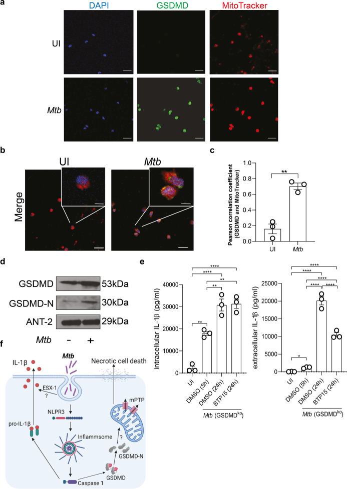
Fig. 6. GSDMD is associated with the mitochondria and Mtb ESX-1 influences IL-1β secretion in infected macrophages.
a, b Immunofluorescence staining of GSDMD in uninfected (UI) and Mtb-infected primary human macrophages. a Cells were stained with DAPI (blue), GSDMD (green), and MitoTracker (red) as indicated. b Merge pictures of a. Images are representative of three individual experiments (scale bar: 50 µM). c Quantification of the Pearson correlation coefficient to determine overlapping signals from the GSDMD and MitoTracker staining from a and b. Standard deviation of the mean is indicated and T test with Welsh’s correction was used to determine statistical significance. **≤0.01. d Detection of GSDMD in mitochondrial lysates of uninfected and Mtb-infected primary human macrophages by western blot analysis. The blot is representative of four individual experiments. e IL-1β concentration (pg/ml) from THP-1 GSDMD knock-out cells infected with Mtb (MOI 1) and treated 5 h post infection with BTP-15 (10 µM), as indicated. IL-1β was measured from total cell lysates and the supernatant at 5 h post infection and 24 h post BTP15 treatment. Results are expressed as mean ± SEM. Analysis was done using one-way ANOVA with Bonferroni post-test (ns, not significant; *≤0.05, **≤0.01, ****p ≤ 0.0001). f Infection of macrophages by Mtb leads to an NLPR3 inflammasome formation and activation with consequent secretion of cleaved IL-1β. Further, Mtb infection induces an accumulation of GSDMD-N at the mitochondria, which is associated with mitochondrial damage MPTP opening. BCL-2, as a mitochondrial protein, interferes with mitochondrial damage. Consequently, Mtb induces necrotic cell death in macrophages.

