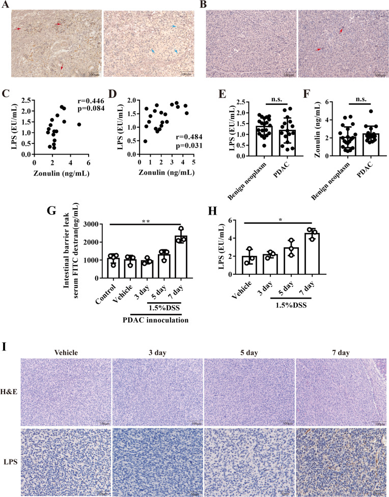
Fig. 1. LPS existed in PDAC tissue and correlated with gut leakage.
A the representative images of immunochemistry staining for LPS staining in pancreatic cancer tissues. B the representative images of immunochemistry staining for LPS staining in PDAC adjacent normal tissues. The correlation between serum LPS and Zonulin in PDAC patients (C) or benign pancreatic neoplasm patients (D). The serum LPS (E) and zonulin level (F) in patients with PDAC or benign pancreatic neoplasm. G FITC conjugated dextran were used to measure intestinal leakage after DSS treatment. H mice serum LPS were measured after DSS treatment. I the typical image of HE and LPS staining in orthotopic PDAC tissue.

