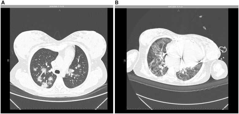
Figure 2.
Non-contrast chest computed tomography scan demonstrated multiple ground-glass opacities associated with thickening of interlobular septal and thin interlaced reticulum, with bilateral multifocal distribution and peripheral and posterior predominance, with extent of lung involvement estimated visually of <50% (A). Nineteen days after a positive COVID-19 test, an increase was observed in multiple ground-glass pulmonary opacities, in addition to sparse foci of consolidation, with bilateral multifocal distribution, the extent of pulmonary involvement, estimated visually to be >50% (B).

